Spotting the Fakes: Understanding the common types of image manipulation in biological research

Images serve as visual evidence in biological research that help researchers validate their experimental findings. They also enable others in the field to reproduce the studies, adding trust and credibility to the findings. However, there has been a concerning rise in image integrity issues, which represent one of the major forms of scientific misconduct. Such issues can lead to serious consequences: reduced citations, retractions, loss of credibility and termination of the researchers involved in the study. Furthermore, this undermines public confidence in research and journals.
To better understand how these issues arise, it is important to first recognize and differentiate acceptable forms of image editing. Generally, any linear adjustments that are made to improve the clarity of the entire image is acceptable. This include:
It is important to note that raw images should always be unaltered. Any necessary modifications should be made only to copies of the original image acquired during the experiments. Journals typically outline these guidelines on their website. Therefore, authors should review the guidelines of their target journal to ensure compliance with image integrity standards.
Common Types of Image Integrity Issues in Biological Research
Duplications
Simple Duplications
In simple duplications, researchers replicate sections of an image without disclosing that duplication has taken place. This can happen when the same region or object within an image, such as a cell or protein band, is copied and pasted elsewhere and is presented as separate data points. Such duplications can happen within the same image panel or across figure panels. This is particularly problematic in studies where the presence or count of specific cells, structures, or protein expressions is crucial.
Example:
In this image, the images in top panel are identical to those in the bottom panel (outlines in red and blue boxes). But, as one can see, the conditions of these two experiments are different, raising a serious question on image integrity of these figure panels.
 Reported in Bik EM, et. al. 2016
Duplications With Repositioning
Duplications with repositioning involves moving, rotating, or reversing parts of an image to create the illusion of distinct and varied data points. This type of manipulation is particularly deceptive, as it falsely suggests a diverse and wide distribution of observed effects. Such practices can leads to incorrect conclusions about the data variability and distribution, ultimately undermining the findings reported in the manuscript.
Example:
In this image, the highlighted sections (outlined in red box) of the western blot images look very similar to each other despite being labeled as different proteins and are presented to be isolated from different cell fractions.
 Reported in Bik EM, et. al. 2016
Alterations
Uneven Changes in Brightness and Contrast
Adjusting the brightness and contrast of a specific part of the image is accounted as image manipulation. This is because selectively increasing the brightness of specific sections of the image can make certain features appear more prominent than they actually are, misleading viewers into drawing incorrect conclusions about the findings.
Example:
In these images on the left panel (before adjustment), the intensity of staining is similar. But, after unequal adjustment of the images, treatment B looks more pronounced than treatment A (which is not true).
Cuts and Beautifications
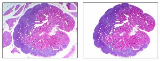
This involves altering the data by removing or modifying certain areas of the image to project a different idea than the one presented by the original image. Unlike acceptable editing practices that enhance image clarity while preserving data integrity, this type of manipulation distorts the raw data in misleading ways, which can significantly affect data interpretation.
Example:
In this image, the background is entirely ‘cleaned up’. This is highly unethical as the image on the right side does not provide information on the uneven exposure and other artifacts that could influence data interpretation.
Smudging and Smearing
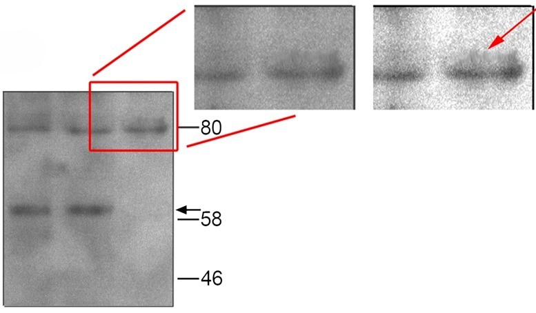
Smudging and Smearing refer to blurring, stretching and/or distorting sections of an image to hide unwanted elements. This is often done to conceal any manipulations made to the original image, making it relatively difficult to detect. This obscures critical information in the image, leading to misinterpretation of the data, ultimately compromising the integrity of the research findings.
Example:
In this image, the presence of a protein band above the 80 kDa band is not visible clearly unless the contrast is modified and gamma correction is performed (indicated by the red arrow). This is a clear example of pixel smearing that is performed to hide the protein band pointed by the red arrow.
 Reported in Blatt M et. al. 2013
Digital Filtering
Digital filtering refers to the use of software filters to enhance the overall image. Digital filtering is acceptable when performed within the scope of ethical guidelines (such as uniform adjustments and filtering to improve the overall clarity of the image) and is reported in the manuscript. However, this can be flagged as unethical when the filtering is performed only to selective regions, leading to data distortion.
Example:
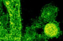
In this example, digital filtering is used to enhance the visual appeal of the graph. However, selective filtering or manipulation of data for visual effect is unethical and is considered unacceptable.
Image created created for demonstration purpose by Enago
Image Fabrication
Image fabrication involves creation or insertion of false data that was never a part of the original experimental result. This kind of manipulation is typically done to add new structures — cells or protein bands and to generate new datasets. This leads to misleading interpretations and loss of credibility of the original findings.
Example 1:
In the below gel images, note how a new band is inserted in lane 3 which was originally absent. This is unethical as this provides a false sense of the presence of a protein (or nucleic acid – DNA or RNA) in the sample which compromises data analysis and interpretation.

Image taken from Rossner et. al. 2004
Example 2:
The microscopic image on the left is misleading because it shows cells from different fields placed next to each other, giving the false impression that they are from the same microscopic field. Here, image manipulation is revealed by adjusting the contrast of the entire image, as shown in the image on the right, which makes the inserted cells visible.
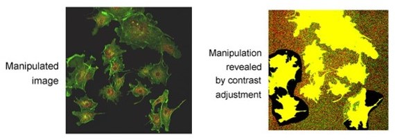
Image taken from Rossner et. al. 2004
Ensuring Image Integrity: A collective effort by all stakeholders
Given the prevalence of these varied forms of image manipulation, it is crucial for journals and institutions to set strict standards and ensure clarity on acceptable image practices. The Committee on Publication Ethics (COPE) has advised journals to provide clear instructions and comprehensive guidelines to authors regarding image manipulation. Most journals have extensive policies in place to keep such image integrity issues in check. Oftentimes, journals mandate the authors to share raw data that will help peer-reviewers cross-verify the authenticity of the images included in the article. They also use image analysis software that help screen the manuscripts systematically for any image integrity issues.
Services that integrate technology with human expertise offer a balanced solution that all the stakeholders of the STM industry can benefit from. By meticulously screening manuscripts with the combined support of advanced software and expert judgement, services like Enago’s image manipulation detection service offer more than just the benefit of detection; they uphold and preserve the values of data integrity which are crucial to the field of research.
On the other hand, effective management of images requires a unified commitment from research groups, institutions, and journals alike. Combined efforts from all stakeholders will help enhance scientific credibility and uphold public trust in the findings uncovered by researchers.
As the academic community continues to evolve with the advent of new technologies and methodologies, the commitment to ethical research practices remains a cornerstone to ensure that the pursuit of knowledge progresses in a manner that is both innovative and honorable.


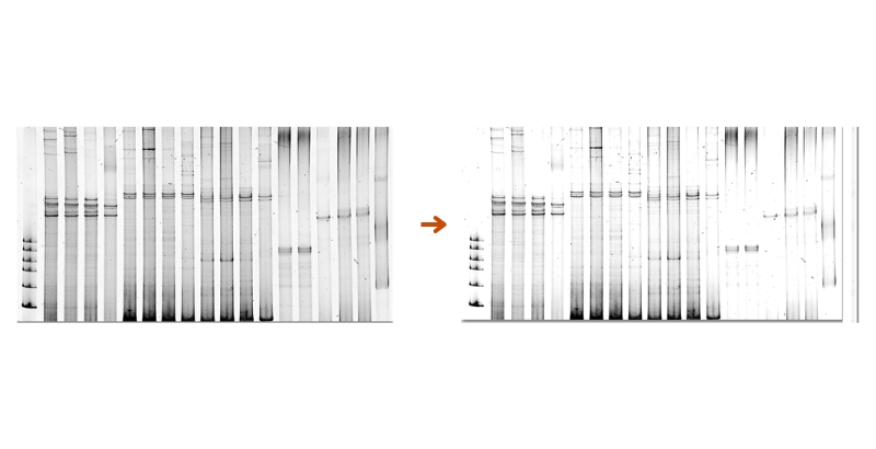 Changes in brightness of the entire image
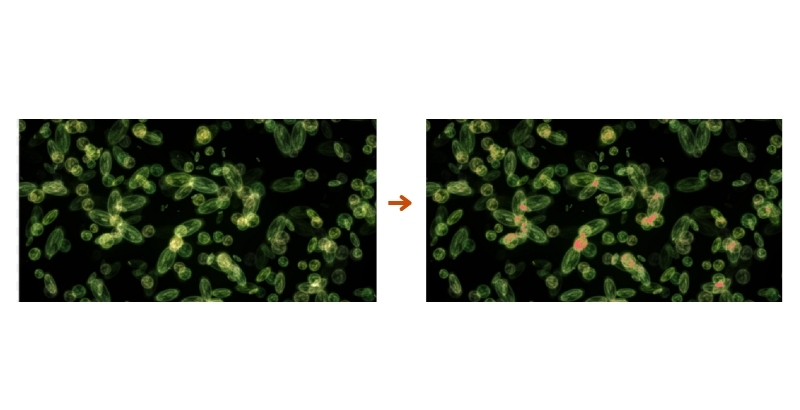 Changes in color of the entire image
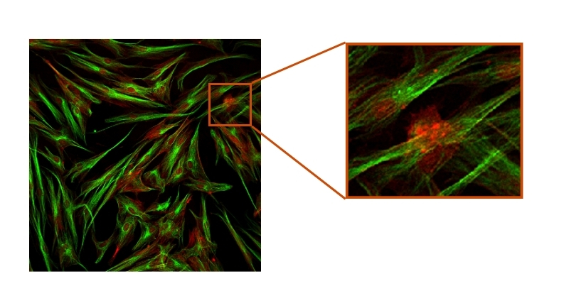 Simple magnification of a section when provided with the original image alongside
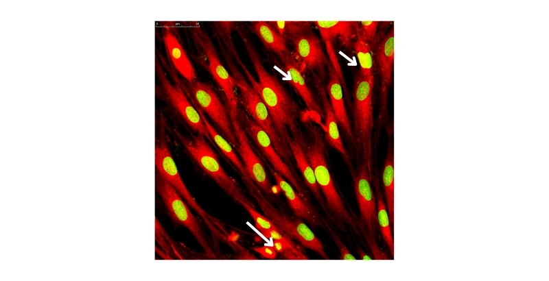 Using arrows and boxes to highlight specific aspects of the image
 Providing scale bars and labels