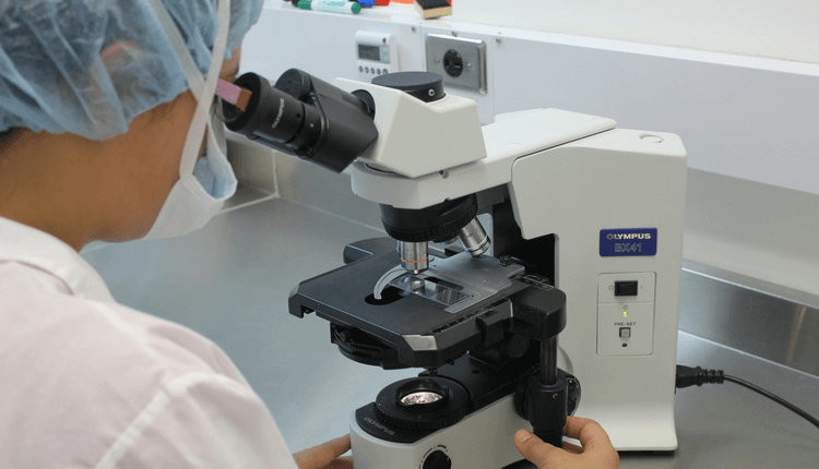
Is It Possible to Get Real-Time Images of Living Organisms?

Until recently, living organisms could only be observed under a microscope, after using a stain. But researchers have now come up with a better procedure. Researchers of the University of Illinois have devised a new microscope system that can take real-time images of living organisms. They have named the technique as “simultaneous label-free autofluorescence multi-harmonic (SLAM) microscopy.” In this technique, researchers use pulses of light that can take simultaneous images at multiple wavelengths. This, in turn, enables researchers to see the different processes occurring simultaneously in living cells. Two major differences between SLAM and other microscopic techniques is that the former uses only living cells and no other chemicals. SLAM will give cancer researchers a new way to visualize the growth and progression of tumor cells.
To know more, click here now!


